
Intraoral Scanner: A Handheld Digital Imaging Device
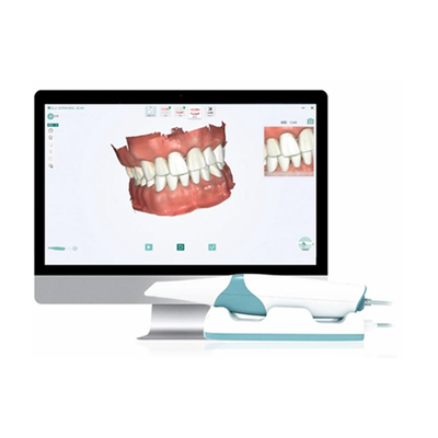
An intraoral scanner is a handheld device designed to directly capture and transfer digital images of the oral cavity to a display screen. The scanner’s light source projects onto scanned objects, such as a full dental arch, and the software processes a real-time 3D model that appears on the monitor.
This device provides high-resolution images, offering detailed visualization of both hard and soft tissues inside the mouth. The scanning process, utilizing visible light or laser beams, takes place chairside within just a few seconds or minutes.
Components of an Intraoral Scanner
- Wired or wireless handheld piece
- Connected to a computer system with a monitor
This technology enhances accuracy, efficiency, and patient comfort in modern digital dentistry.
How Intraoral Scanners Work
An intraoral scanner consists of a handheld scanning camera, a computer, and specialized software. The scanner connects to a computer running the intraoral scanning software, which processes the digital data captured by the camera. The smaller the scanner tip, the more flexible and precise it is in reaching deeper areas of the oral cavity for accurate data collection.
Scanning Process
- Positioning the Scanner: The dentist carefully places the intraoral scanner’s head inside the patient’s mouth.
- Capturing Images: The scanner moves smoothly across the tooth surfaces, automatically recording the size and shape of each tooth.
- Processing the Scan: Within one to two minutes, the system generates a detailed digital image.
- Real-time Visualization: The dentist can view, zoom in, and edit the images on the computer screen for better analysis.
- Data Transmission: The digital scan is sent to a dental laboratory for fabricating custom restorations.
This efficient process saves time, improves accuracy, and allows dentists to treat more patients in a shorter period.
History and Development of Intraoral Scanners
In the 18th century, dentists used materials like Impregum, dense silicone, and alginate for impressions. However, these traditional methods were inaccurate, uncomfortable, and time-consuming.
To overcome these limitations, intraoral scanners emerged as a digital alternative. This innovation coincided with the development of CAD/CAM technology:
- 1970s: The concept of computer-aided design (CAD/CAM) was introduced to dentistry by Dr. François Duret.
- 1985: The first commercially available intraoral scanner was introduced and used in laboratories to create precise restorations.
- Modern Advancements: Today’s scanners are smaller, faster, and more accurate, revolutionizing digital dentistry with efficient workflows and high-precision treatment planning.
Dentists increasingly adopt digital dentistry due to its accuracy, efficiency, and enhanced patient experience.
Important considerations for using intraoral scanners
To achieve an accurate intraoral scan, undercuts should be avoided, and the patient’s bleeding must have stopped. Some systems require additional preparation by covering the scanning area with a thin layer of powder. Implant platforms are scanned indirectly by placing a “scan body.” When preparing the teeth for intraoral scanning, parameters such as the distance between teeth and the opposing teeth, the placement path, alignment of the supporting teeth, and the morphology of the tooth surface are crucial. A full digital jaw scan is created by merging the scanned images of both the upper and lower jaws. Specific areas can be easily rescanned and combined for better precision.
Key Advantages of Intraoral Scanners
1. Reduced Material Waste
- Eliminates the need for PVS materials, plaster, and impression trays.
- Saves on consumable costs and minimizes material wastage.
2. Faster Diagnosis & Treatment
- Real-time visualization accelerates treatment planning and decision-making.
3. Higher Diagnostic Accuracy
- Digital scans allow precise image magnification, editing, and multi-angle analysis.
- Eliminates distortions common in traditional impressions.
4. Digital Storage & Easy Retrieval
- Patient scans can be stored in cloud storage or internal databases.
- Easily accessible for future reference, sharing with labs or other specialists.
5. 3D Model Processing for Restorations
- Digital scans are saved in STL format, compatible with CAD/CAM software.
- Used for implant placement, crown fabrication, and bite analysis.
- Reduces the need for multiple patient visits.
6. Improved Patient Comfort
- No need for traditional impression materials, reducing gag reflex and discomfort.
- Shorter scan times make the procedure quick and efficient.